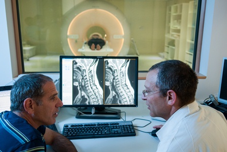
Above: Prof. Alon Friedman (left) and Dr. Ilan Shelef with the MRI machine
A team of brain scientists tested former Israeli prime minister Ariel Sharon late last week using functional MRI to assess his brain responses. The team consisted of Prof. Alon Friedman, and Drs. Galia Avidan and Tzvi Ganel of BGU's Zlotowski Center for Neuroscience. Erez Freud, a PhD student in the Department of Psychology, Dr. Ilan Shelef, head of Medical Imaging at Soroka University Medical Center, and Professor Martin Monti, from the Departments of Psychology and Neurosurgery at the University of California Los Angeles.
Ariel Sharon, presumed to be in a vegetative state since 2006 due to brain hemorrhage, was scanned to assess the extent and quality of his brain processing using methods recently developed by Professor Monti and collaborators.
During the test, which lasted approximately two hours, scientists showed Mr. Sharon pictures of his family, made him listen to his son’s voice, and used tactile stimulation to assess to what extent his brain responded to external stimuli. To their surprise, significant brain activity was observed in each test in specific brain regions, indicating appropriate processing of these stimulations.
Additional tests were performed to assess his level of consciousness. While there were some encouraging signs, these were subtle and not as strong. Monti said that “Information from the external world is being transferred to the appropriate parts of Mr. Sharon’s brain, however, the evidence does not as clearly indicate whether Mr. Sharon is consciously perceiving this information.”
Shelef mentioned that “this kind of forefront research was a main motivation for the recent acquisition of a state of the art MRI machine by BGU and Soroka Medical Center.”
Ganel, who initiated the project, stressed the wish of Sharon’s family to employ these techniques not only for the benefit of the former prime minister, but also for other families in similar situations.
Indeed, according to Friedman, head of the Zlotowski Center for Neuroscience, “It is important that these new techniques be available in Israel for the large number of patients considered to be in a Vegetative State. Knowing what sensory channels are intact in these patients is crucial for the family and the treating team to stimulate and interact with them.”
Avidan stated that “This line of research could shed light on basic questions pertaining to the neural basis of consciousness.”
The MRI is a Philips INGENIA 3T and is part of a joint project by BGU and Soroka to further brain research and medical assessment. Funding for the purchase came through the efforts of the American Associates, BGU.