Dr. Netta Vidavsky, the newest member of BGU's Department of Chemical Engineering, studies breast microcalcifications, or the small particles detected in mammography that are used as early diagnostic markers for the disease. To be exact, she studies those calcifications' crystal structures, which have increasingly been linked with the cancer's pathology. By going back to the very beginnings of the tumor's development — even before, that is, it was visible to the human eye — Vidavsky is finding clues to its progression, with implications for the future of breast-cancer prognostics, diagnostics, and therapy.
“It's critical that we continue to develop new treatments for breast cancer, with the goal of improving patient outcomes," Vidavsky says. “But it's equally critical to determine who does and does not need these advanced treatments, which may be effective, but can also have serious side effects," both in terms of the health of the patient and the financial costs to our healthcare system. If Vidavsky can show that certain calcifications' compositions make them unlikely candidates for invasive cancer, her research can not only help medicine's efforts to personalize the approach to treatment. It can also, she believes, lead to the inhibition of the formation of those calcifications in the first place — and of the cancer associated with their unchecked growth.
When asked why these microcalcifications haven't been the subject of more study, Vidavsky muses that it takes a chemist—or at least, “someone less interested in interpreting the calcifications' macroscopic patterns or groupings on a mammogram than in characterizing their composition, structure, and properties." She traces her fascination with the latter to her work at a surface-chemistry factory in the years following her undergraduate degree. “My job was to extract lycopene crystals from tomatoes for use as a natural food coloring. But I was more interested in the lycopene crystal's structure than in improving its stability in emulsions," she recalls. That fascination led to her graduate studies at the Weizmann Institute of Science, where she focused on how minerals form skeletons in biological systems, and later, during her postdoctoral studies at Cornell University, on why they sometimes grow out of control in disease.

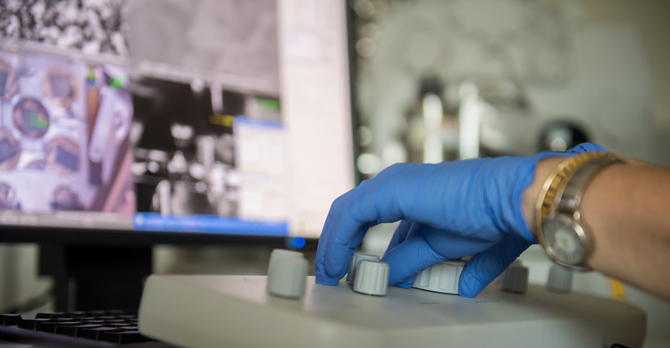
Of course, there is also the issue of size, or in the case of microcalcifications, the lack thereof. As the name suggests, most calcifications are too small to observe properly using low-resolution mammography. They are also, like all biological samples, found in tissues that contain large amounts of water, which presents a challenge all its own. “Most advanced microscopes — the ones that allow us to see at the nanoscale — use a vacuum. But since water evaporates immediately in a vacuum, we would first need to take the water out of the sample, and that would change the structure of the tissue itself." The answer? “High-pressure freezing," says Vidavsky. “It immobilizes all the molecular components of a sample within milliseconds, allowing us to preserve and then study their structure in a high-vacuum cryogenic scanning electron microscope."
Fortunately, Ben-Gurion University's Ilse Katz Institute for Nanoscale Science and Technology has both the microscope and the high-pressure freezing device—a combination, Vidavsky points out, not to be taken for granted. While conducting her postdoctoral studies in materials science and engineering in New York, Vidavsky would need to bring her samples to a nearby institution that did have high-pressure freezer: Northwestern University, in Evanston, Illinois.
The benefits of proximity apply to more than just the equipment, however. “One of the great things about working at Ben-Gurion University is its relationship with the adjacent Soroka University Medical Center, with whose doctors I work very closely," Vidavsky explains. “Not only do SUMC's pathologists and endocrinologists provide me with clinical samples for my research, but they also play an important role in helping me understand what I'm seeing. We constantly consult with each other, sharing questions and insights that can advance both our goals."
This collaborative approach is reflected in the increasingly cross-disciplinary nature of science, which Ben-Gurion University — home to some 60 different interdisciplinary centers — actively encourages among its researchers and seeks out in new faculty recruits. Indeed, if there was a time when chemists might not have felt at home in the cancer-research community, today many of the innovative approaches to cancer detection, prevention, and treatment are coming from scientists such as Vidavsky, who combine elements of chemical and biomedical engineering with materials science to come at the challenge from new angles. For Vidavsky, this development is great for cancer research and for chemistry, too.
“In my Introduction to Chemical Engineering course, I always begin by asking my students who's here because he or she loves chemistry but is afraid it will be too hard to find a job without the engineering. Nearly 80 percent of students raise their hands," Vidavsky says. “I make sure to warn them that chemical engineering does mean a lot of engineering. Fortunately, I can also reassure them that there's more and more room in fields such as cancer research for chemical engineers, even ones who are chemists at heart. I hope this encourages more talented students and researchers to pursue careers at the intersection of different disciplines, for the benefit of both science and society."